

Ultrasound
It is a modern stethoscope of present doctor most commonly and most widely used without any hazard of radiation and thus most useful and popular in obstetrics Sonography that is for pregnant mothers and their unborn baby. At present colored ultrasound machine are in routine use which facilitates to watch blood flow by color Doppler technique which is based on a very well known scientific effect called Doppler’s effect. Doppler is used in all body parts except lungs and bones.
It is most commonly used for abdominal problem and pregnancy related scanning.
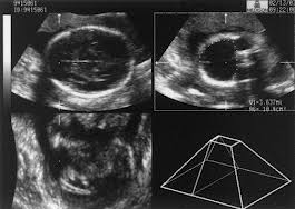
Use of Ultrasound in Pregnancy- A minimum of three scan are required during normal pregnancy
1st in the 11th to 13th week for dating of pregnancy particularly when screening is required for down syndrome and other genetic problem.
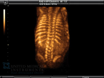
2nd in the 18th to 22nd week for detail study of the baby whether developing normally or not which is called level 2 scan
3rd in the 28th to 40th week for growth parameter.
Another scan preferably at the time of delivery is recommended.
Ultrasound Preparation
It is preferable to come empty stomach for 6 hours for upper abdominal problem that is gall bladder, liver and kidneys.
Urinary bladder should be full so that first trimester pregnancy and urinary bladder pathology can be imagined otherwise you will be requested to wait till bladder is full.
Procedure time- 10-30 mins (depends on bladder full and condition of patient)
Other important uses of Ultrasound are-
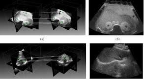
Echocardiography- which is available for pediatric, fetal (unborn) and adult patient it is to see the size and function of various valves and valve in it and with the advent of color Doppler the function of the heart can be assessed properly particularly if suspecting heart problem.
Special use of Ultrasound in breast
It is done on breast for any mass or any suspected or confirmed mass and to supplement breast cancer screening along with mammography. Breast tissue biopsy is done under guidance of ultrasound.
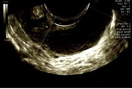
Color flow ultrasonography (Doppler) of a carotid artery - scanner and screen

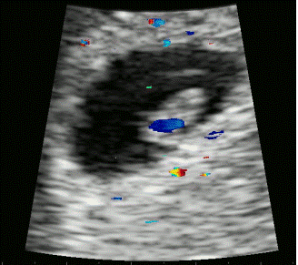
Doppler ultrasound - Images of fetal heart (heart beat)
TIME DURATION OF AN ULTRASOUND SCAN
The majority of scans take between 10 to 30 minutes and will usually occur in the Radiology department in a Diagnostic Center.
 |
 |
 |
||
 |
 |
 |
||
 |
 |
 |
||
 |
 |
 |
||
 |
 |
|||